

Translate this page into:
Giant Retroperitoneal Sarcoma: A Case Report and Review of Literature
This article was originally published by Thieme Medical and Scientific Publishers Private Ltd. and was migrated to Scientific Scholar after the change of Publisher.
Abstract
Liposarcomas are neoplasms of mesodermal origin derived from adipose tissue representing 10 to 14% of all soft tissue sarcomas, with the most frequent subtype being liposarcoma. Given that the retroperitoneum is a large space in which the retroperitoneal liposarcoma can grow asymptomatically up until a mass effect develop. Hence, patients usually present late with symptoms with possible invasion to nearby structures. These tumors are known to reach significantly large dimension, despite their poor vascularization and can grow to enormous size, the average diameter of the tumor is 20 to 25 cm with a weight of 15 to 20 kg. Surgery with en-bloc resection of the tumor and adherent nearby structures, with intact capsule, remains the gold standard in surgical management of retroperitoneal sarcomas. Herein, we present a case of 52 year old male patient with a huge 48 cm right-sided retroperitoneal liposarcoma, managed surgically by en bloc excision of the tumor, right kidney, right ureter, right adrenal gland, and the right colon.
Keywords
sarcoma
sarcoma retroperitoneal
sarcoma retroperitoneal

Introduction
Liposarcomas are neoplasms of mesodermal origin derived from adipose tissue representing 10 to 14% of all soft tissue sarcomas, with the most frequent subtype being liposarcoma (41%).1 Retroperitoneal liposarcomas comprise 0.07 to 0.2% of all neoplasia.2 Approximately 85% of these are malignant, liposarcoma is the most frequent histopathological variant of the retroperitoneal sarcomas.3 In view that the retroperitoneum is a large space in which the retroperitoneal liposarcoma can grow asymptomatically up until a mass effect develop. Hence, patients usually present late with symptoms like vague abdominal discomfort, increasing abdominal girth and lump abdomen. Diagnosis is usually made on the basis of computed tomography (CT) scans of the abdomen and pelvis. Surgery with en bloc resection of the tumor and adherent nearby structures is the gold standard of treatment. Herein, we present a case of 52-year-old male patient with a huge 48-cm right-sided retroperitoneal liposarcoma, managed surgically by en bloc excision of the tumor, right kidney, right ureter, right adrenal gland, and the right colon.
Case Presentation
A 52-year-old male patient, previously healthy with no previous abdominal surgeries or any remarkable family history, presenting with the complaint of increased abdominal girth, 6 months prior to presentation. Patient denied any weight loss, fatigue, and change bowel habits. On physical exam, his abdomen was markedly swollen and felt hard without any tenderness. Blood workup including a complete blood count, electrolytes, creatinine, and liver function tests done and turned out to be normal. Hence, a colonoscopy and CT scan abdomen pelvis with intravenous (IV) contrast were scheduled. Colonoscopy was normal. However the CT scan showed solid mass located in the right abdomen, with its central region not absorbing the intravenous contrast, which was attributed to central necrosis or cystic degeneration. The mass occupied the right portion of the abdomen displacing the right colon, with its upper borders to the lower edge and of the liver and lower borders reaching the pelvis (►Figs. 1 and 2) and in close proximity to the rectosigmoid colon and compressing the bladder. The size of this mass was measured to be 48 cm × 31 cm × 12 cm. The position of the mass was displacing the anatomical structures and organs away from their natural position. In addition, the tumor was in contact with the right kidney, the inferior vena cava and the right renal vein causing mild dilation of the right pelvis of the kidney. Patient was scheduled for surgical excision of the mass with an R0 resection as the aim. Surgery performed with a generous midline incision from the xiphoid process to the pubic bone with extension to the right subcostal area (►Fig. 3). The tumor was exposed, displacing the right colon past the midline, with no clear cleavage plane in between. We opted for en bloc resection of the tumor, right adrenal, right kidney, and the right colon (►Fig. 4). Pathology turned out to be well differentiated liposarcoma. Patient had a smooth postoperative course and was discharged home day 4 after surgery. On 2-year follow up, the patient is disease free as documented by imaging with no complaint.
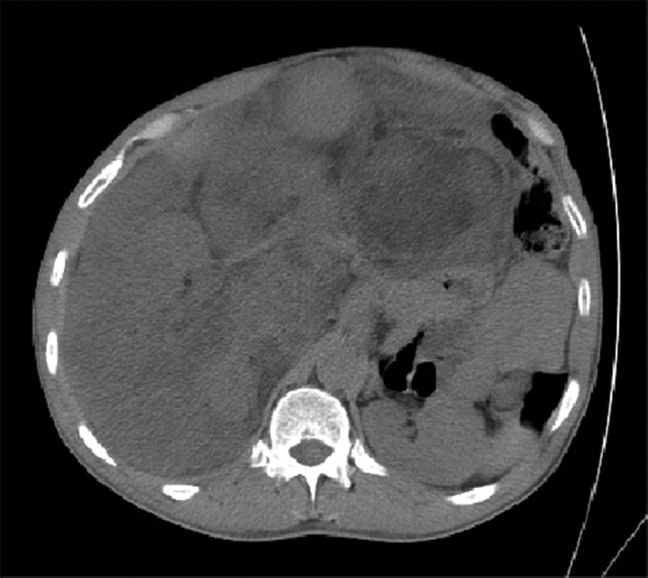
- CT scan showing the large retroperitoneal sarcoma with abdominal structures being displaced to the left. CT, computed tomography.
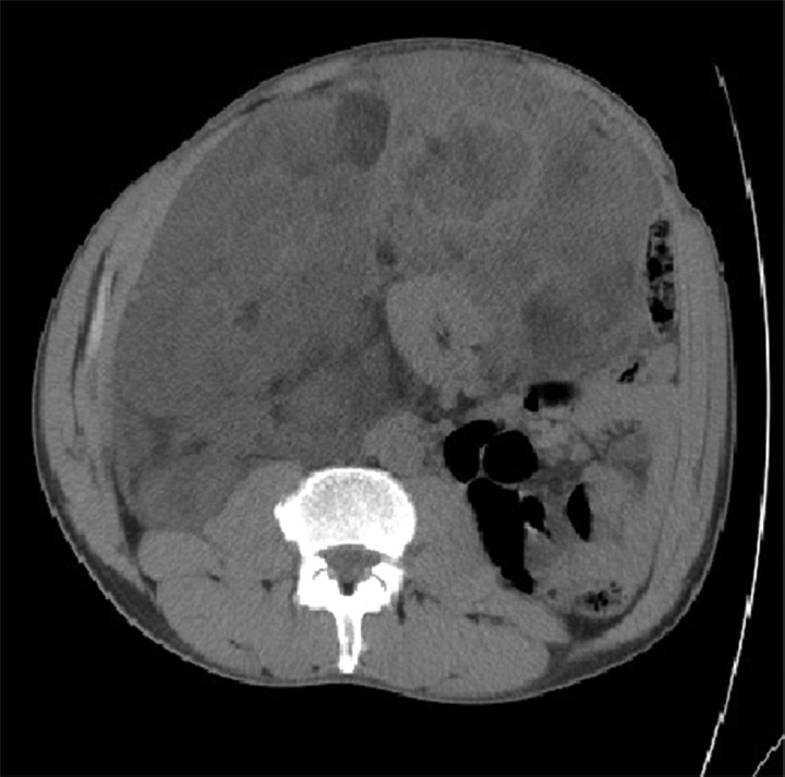
- CT scan showing the large retroperitoneal sarcoma. CT, computed tomography.

- Intraoperative picture showing the exposed tumor after the midline incision being extended to the right subcostal.

- Tumor with kidney, ureter and the right colon en bloc excision.
Discussion
In view that retroperitoneal liposarcomas can grow asymptomatically up until a mass effect starts to appear. These tumors are known to reach significantly large dimension, despite their poor vascularization, and can grow to enormous size, the average diameter of the tumor is 20 to 25 cm with a weight of 15 to 20 kg.2 Surgery with en bloc resection of the tumor and adherent nearby structures, with intact capsule, remains the gold standard in surgical management of retroperitoneal sarcomas. Moreover, only a complete surgery has been proven to improve the overall survival in patients diagnosed with retroperitoneal sarcomas, with the most important variable being en bloc resection of the tumor with adherent nearby structures. Aforementioned, studies have shown that the median survival of patients with complete resection was 103 months versus 18 months with incomplete resection, similar to when no surgery was done at all.1 For tumors more than 10 cm in dimensions, complete R0 resection can be achieved in 70% of cases; however, multiorgan resection is deemed necessary in 50% of cases to achieve R0 resection.3 Hence, the resection of a retroperitoneal sarcoma of remarkable size is a challenge due to multiple factors, the anatomical site, absence of an anatomically evident vascular, and lymphatic pedicles making it hard to obtain safe margin, adherences to nearby organs, and the great vessels. In our case, the tumor was among the largest reported in literature (►Table 1) adherent to the right kidney and right colon, hence curative surgery necessitates the en bloc excision of the right retroperitoneal tumor, right adrenal gland, right kidney, right ureter, and the right colon. Giant retroperitoneal sarcomas has been rarely reported in the medical literature. To our knowledge, 20 cases has been reported with tumor size 40 cm in greater dimension and above (►Table 1), with a mean age of presentation being 53 years old, the female-to-male ratio is 8:12, 50% requiring multiorgan resection and the rate of R0 resection was 100%. Following surgical resection, 50 to 100% of liposarcomas recur from residual tissue, which is the primary cause of death.4 Therefore an aggressive surgical behavior is mandated, with the resection of the structures and viscera adjacent to the pathological process in an attempt to obtain an R0 resection. The use of chemotherapy is limited to metastatic disease as palliation, along with radiotherapy in nonoperable cases or incomplete resection.5,6 In summary, the ability to achieve R0 resection along with the histopathologic grade will solely determine the disease prognosis. The abovementioned two factors are determined during surgery and are very hard to elucidate preoperatively.
| No. | Author (y) | Gender/age (y) | Preimaging | Size (cm3) | Histological subtype | Complete resection | Multiorgan resection |
|---|---|---|---|---|---|---|---|
| 1 | Herrera-Gómez et al7 (2008) | M/24 | CT | 80 × 50 × 35 | Undifferentiated | Yes (R0) | No |
| 2 | Zeng et al8 (2017) | M/45 | CT | 65 × 45 × 30 | Well-differentiated | Yes (R0) | No |
| 3 | Benseler et al9 (2009) | M/39 | CT | 60 × 50 × 36 | Well-differentiated | Yes (R0) | Yes |
| 4 | Hazen and Cocieru10 (2017) | M/64 | CT | 60 × 42 × 31 | Poorly differentiated | Yes (R0) | No |
| 5 | Akhoondinasab et al11 (2011) | M/54 | US /CT | 58 × 45 × 36 | Well-differentiated | Yes (R0) | No |
| 6 | De Nardi et al12 (2012) | M/40 | CT | 50 × 49 × 35 | Well-differentiated | Yes (R0) | No |
| 7 | Yol et al13 (1998) | M/63 | US/CT | 50 × 45 × 32 | Myxoid | Yes (R0) | Yes |
| 8 | Selmani et al14 (2011) | F/58 | US/CT | 50 × 35 × 25 | Well-differentiated | Yes (R0) | No |
| 9 | Our Case | M/52 | CT | 48 × 31 × 12 | Well-differentiated | Yes (R0) | Yes |
| 10 | Clar et al15 (2009) | M/66 | CT | 47 × 42 × 25 | Well-differentiated | Yes (R0) | Yes |
| 11 | Sharma et al16 (2013) | F/60 | CT | 47 × 40 × 25 | Well-differentiated | Yes (R0) | No |
| 12 | Morandeira et al17 (2008) | F/63 | CT | 45 × 43 × 24 | Myxoid | Yes (R0) | Yes |
| 13 | Hashimoto et al18 (2010) | M/41 | CT | 45 × 40 × 30 | Poorly differentiated | Yes (R0) | Yes |
| 14 | Oh et al19 (2016) | F/71 | US/CT | 45 × 30 × 11 | Well-differentiated | Yes (R0) | No |
| 15 | Xie et al20 (2006) | F/41 | US/CT | 43 × 40 × 25 | Well-differentiated | Yes (R0) | No |
| 16 | Caizzone et al21 (2015) | F/64 | CT | 42 × 37 × 18 | Mixed | Yes (R0) | Yes |
| 17 | Bansal et al22 (2013) | M/52 | CT | 40 × 35 × 35 | Mixed | Yes (R0) | Yes |
| 18 | Liu et al23 (2013) | F/55 | CT | 40 × 30 × 20 | Well-differentiated | Yes (R0) | No |
| 19 | Zheng et al24 (2011) | M/55 | CT | 40 × 30 × 20 | NA | Yes (R0) | Yes |
| 20 | Salemis et al25 (2009) | F/54 | CT/MRI | 40 × 26 × 16 | Mixed | Yes (R0) | Yes |
| 21 | Fu Qiang26 (2007) | M/52 | CT | 40 × 30 × 10 | Well-differentiated | Yes (R0) | Yes |
Abbreviations: CT, computed tomography; F, female; M, male, MRI, magnetic resonance imaging; US, ultrasound.
Conclusion
Retroperitoneal liposarcomas are a unique disease and require an aggressive surgical approach with multiorgan resection if necessary, in accordance with the ability of the patient to tolerate the procedure. Surgery with R0 resection remains the cornerstone in its management, with chemotherapy and radiotherapy reserved for palliation.
Conflict of Interest
None declared.
References
- Retroperitoneal soft-tissue sarcoma: analysis of 500 patients treated and followed at a single institution. Ann Surg. 1998;228(03):355-365.
- [CrossRef] [PubMed] [Google Scholar]
- [Giant retroperitoneal liposarcoma] (in Spanish) Cir Esp. 2005;77(05):293-295.
- [CrossRef] [PubMed] [Google Scholar]
- Operative management of primary retroperitoneal sarcomas: a reappraisal of an institutional experience. Ann Surg. 2004;239(02):244-250.
- [CrossRef] [PubMed] [Google Scholar]
- Giant recurrent retroperitoneal liposarcoma initially presenting as inguinal hernia: Review of literature. Int J Surg Case Rep. 2012;3(03):103-106.
- [CrossRef] [PubMed] [Google Scholar]
- Retroperitoneal tumors. Retroperitoneal myxoid liposarcoma. Report of a new case.] (in Spanish) Arch Esp Urol. 2000;53(02):170-173.
- [Google Scholar]
- [Adipose retroperitoneal tumors. Apropos a giant myxoid liposarcoma.] (in Spanish) Actas Urol Esp. 1996;20(01):79-84.
- [Google Scholar]
- Giant retroperitoneal liposarcoma. World J Surg Oncol. 2008;6:115.
- [CrossRef] [PubMed] [Google Scholar]
- Clinicopathological characteristics and experience in the treatment of giant retroperitoneal liposarcoma: a case report and review of the literature. Cancer Biol Ther. 2017;18(09):660-665.
- [CrossRef] [PubMed] [Google Scholar]
- [Case report–surgical therapy of a retroperitoneal liposarcoma weighing 45 Kg.] (in German) Zentralbl Chir. 2009;134(02):174-177.
- [CrossRef] [PubMed] [Google Scholar]
- Giant retroperitoneal sarcoma. J Gastrointest Surg. 2017;21(03):602-603.
- [CrossRef] [PubMed] [Google Scholar]
- Recurrent giant liposarcoma of the spermatic cord. Urology. 2012;79(01):113-114.
- [CrossRef] [PubMed] [Google Scholar]
- Retroperitoneal and scrotal giant liposarcoma: report of a case. Surg Today. 1998;28(03):339-342.
- [CrossRef] [PubMed] [Google Scholar]
- Giant retroperitoneal liposarcoma: a case report. Prilozi / Makedonska akademija na naukite i umetnostite, Oddelenie za biološki i medicinski nauki = Contributions/Macedonian Academy of Sciences and Arts. Section Biol Med Sci,. 2011;32:323-332.
- [Google Scholar]
- Interdisciplinary resection of a giant retroperitoneal liposarcoma of 25 kg. ANZ J Surg. 2009;79(12):957.
- [CrossRef] [PubMed] [Google Scholar]
- Giant inflammatory variant of well differentiated liposarcoma: a case report of a rare entity. J Clin Diagn Res. 2013;7(08):1720-1721.
- [CrossRef] [PubMed] [Google Scholar]
- Surgical treatment of a giant liposarcoma in a Japanese man. Adv Urol. 2010;2010:943073.
- [CrossRef] [PubMed] [Google Scholar]
- A giant retroperitoneal liposarcoma encasing the entire left kidney and adherent to adjacent structures: a case report. Case Rep Oncol. 2016;9(02):368-372.
- [CrossRef] [PubMed] [Google Scholar]
- Giant retroperitoneal liposarcoma: experiences in diagnosis and treatment of two cases. Guizhou Med J. 2006;30:30-31.
- [Google Scholar]
- Giant retroperitoneal liposarcoma: Case report and review of the literature. Int J Surg Case Rep. 2015;9:23-26.
- [CrossRef] [PubMed] [Google Scholar]
- Giant retroperitoneal liposarcoma- renal salvage by autotransplantation. Indian J Surg. 2013;75(02):159-161.
- [CrossRef] [PubMed] [Google Scholar]
- Giant abdominal liposarcoma: a case report. Zhongguo Laonianxue Zazhi. 2013;33:452.
- [Google Scholar]
- Giant retroperitoneal liposarcoma with mixed histological pattern: a rare presentation and literature review. J Gastrointest Cancer. 2009;40(3,4):138-141.
- [CrossRef] [PubMed] [Google Scholar]
- Huge retroperitoneal liposarcoma: a case report. Chin Med J (Engl). 2007;120(12):1117-1118.
- [CrossRef] [PubMed] [Google Scholar]


