Translate this page into:
Descriptive and Surgical Analysis of 196 Cases of Traumatic Maxillofacial Fractures: An experience of 6 years
*Corresponding author: Narendra S. Mashalkar, MBBS, MS, Mch, Department of Plastic Surgery and Burns, St John’s Medical College, Bengaluru, Karnataka, 560034, India. plasticnaren2005@yahoo.co.in
How to cite this article: Mashalkar NS, Shetty N, Ellur S. Descriptive and Surgical Analysis of 196 Cases of Traumatic Maxillofacial Fractures: An Experience of 6 years. Int J Recent Surg Med Sci. 2024;10:S44-9. doi: 10.1055/s-0043-1761506
Abstract
Objectives
To analyze the etiology, anatomical pattern, and management of upper, midface, and lower face fractures pertaining to our demography and compare our results with other regions and worldwide.
Material and Methods
A 6-year retrospective record analysis from 2013 to 2018 of eligible patients’ data was recorded with a prepared proforma. Demographic parameters including age, sex, etiology, anatomical site, closed or open, displaced or un displaced fracture, type of treatment, associated with head injury, and implants used were evaluated.
Inclusion criteria were all patients with facial bone fractures irrespective of age and gender.
Exclusion criteria were patients with pure soft tissue injury of the face and with facial burns.
Results
Most were involved with multiple facial bone fractures. Out of 196, 72 patients (pts) had involvement of mandible fractures, 79 had involvement of the maxilla, 65 zygoma, 68 nasal bone, 42 orbital wall, 21 frontal bone with processes, and 7 NOE involvement. The most frequent etiologic factor was detected to be road traffic accidents (RTA; 162 ,83%), due to falling (24, 12%), and assault (10, 5%).
In total, 173 were male (88%) and the rest 23 were female (12%). The mean age was found to be 29 years. Twelve patients (6.1%) were less than 14 years of age. Most RTAs had occurred in young adults from 16 to 30 years of age group. We analyzed individual bone fracture involvement and compared it with other geographical locations.
Conclusions
Most facial fractures are combined involving multiple bones in young adults with RTA as the most common etiology. There was a balance seen in managing the facial fractures between conservative and operative methods. These data provide us the information in evaluation of the preventive measures to be taken and give the direction of focusing the clinical and research priority in the future.
Keywords
Facial fractures
Demography
Surgical analysis
INTRODUCTION
Maxillofacial fractures are one of the most important health problems and they are often associated with severe functional and cosmetic morbidity.[1] Facial fractures vary from region to region because of many factors, of which, social, cultural, and environmental factors are important.[2]
Alteration of facial features of an individual may have functional, psychological, social, and professional impact.[3] Analyzing the pattern and epidemiological factors of injuries in specific geography provides data for implementing adequate prevention, diagnosis, and treatment.[3] We have not come across any demographic data in our region pertaining to facial fractures. Facial fractures also are accompanied by other co-existing injures. They must be identified early as some of them are life-threatening.[3,4]
Hence, our main objective was to understand the pattern of facial fractures presenting to us and analysis of the current management of maxilla-facial fractures, particularly mandible fractures being done at our center. We also intend to compare our pattern and surgical analysis to other regions so that an optimal treatment may be given to patients with a scope to excel in our management and give better quality of life in terms of functional and cosmetic appearance to our patients.
MATERIAL AND METHODS
A 6-year retrospective record analysis from 2013 to 2018 of eligible patients’ data was performed with a prepared proforma. Institutional ethics clearance was obtained, and data were tabulated in the form of figures and tables. Demographic parameters including age, sex, etiology, anatomical site, closed or open, displaced or undisplaced fracture, type of treatment, associated with head injury, and implants used were evaluated.
Inclusion criteria: All patients with facial bone fractures irrespective of age and gender were included.
Exclusion: Patients with pure soft tissue injury of the face and with facial burns were excluded.
RESULTS
Most maxillo-facial fractures had an involvement of multiple facial bones.
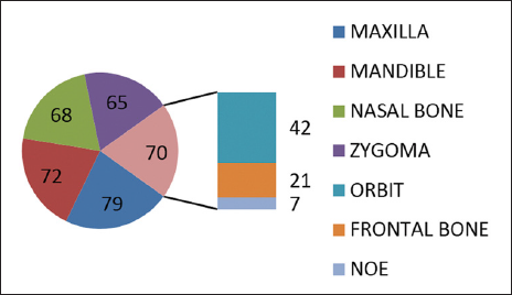
We analyzed the facial bone fractures according to individual bones and described them in the same manner. We recorded a total of 196 facial bone fractures in a 6-year period, which satisfied inclusion criteria into the study category. We excluded many facial bone fractures due to inadequate data. Out of 196, 72 patients (pts) had involvement of mandible fractures, 79 had involvement of the maxilla, 65 zygoma, 68 nasal bone, 42 orbital wall, 21 frontal bone with processes, 7 NOE involvement [Table 1, Figure 1]. The most frequent etiologic factor was detected to be road traffic accidents (RTAs) 162, due to falling (24, 12%), and assault (10, 5%).
Anatomical site involvement |
Number |
|---|---|
Maxilla |
79 |
Mandible |
72 |
Nasal bone |
68 |
Zygoma |
65 |
Orbit |
42 |
Frontal bone |
21 |
NOE |
07 |
NOE, naso-orbito ethmoidal

- Pie chart depicting maxillofacial fracture involvement. NOE, naso-orbito ethmoidal
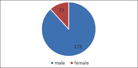
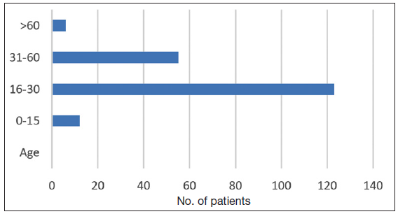
Also, 173 were male (88%) and the rest 23 were female (12%) [Table 2, Figure 2]. The mean age was found to be 29 years. Twelve patients (6.1%) were less than 14 years of age. Most RTAs had occurred in young adults in the 16 to 30 years of age group [Table 3, Figure 3].
Gender |
Number |
|---|---|
Male |
173 |
Female |
23 |
Total |
196 |

- Pie chart depicting gender distribution.
Age (y) |
Number |
|---|---|
0–15 |
12 |
16–30 |
123 |
31–60 |
55 |
>60 |
06 |
Total |
196 |

- Bar diagram depicting age distribution.
Maxillary fracture involvement: Most injuries were compound fractures (71, 90%) and (8, 10%) were closed injuries. Of 79 cases, 14 (18%) were isolated and 65 (82%) were combined.
Most maxilla fractures had an involvement of anterior and lateral walls.
Fifty (63%) patients of fracture of the maxilla underwent surgery due to displacement and functional deformity, and 29 (37%) had conservative management.
Orbit fractures
Of 42 cases, 11 (26%) were isolated and 31 (74%) were combined.
The most common involvement was of the floor (31, 73%), followed by lateral wall (13), 9 had medial wall, 24 inferior orbital margin, 7 had superior orbital margin with extension into the roof. Sixteen (38%) patients of fracture of the orbit underwent surgery due to the displacement and functional deformity. Twenty-six (62%) patients had conservative management.
Frontal bone
We had 21 cases of frontal bone fractures. All injuries were compound and combined with other facial fractures, except one which was closed.
Naso-orbito ethmoidal
Of 7 cases majority were compound combined fractures. 2[28%] fractures of naso-orbito ethmoidal (NOE) underwent surgery due to displacement and functional deformity (5, 82%) had conservative management.
Nasal bone fracture
We had 68 cases of nasal bone fractures, 59 (91%) injuries were compound fractures except 9 (9%) which were closed. Of 68 cases, 44 (68%) were isolated and 24 (32%) were combined.
Forty-one (60%) fractures underwent nasal bone reduction surgery due to displacement and functional deformity, 27 (40%) had conservative management.
Zygoma
We had 65 cases of zygoma bone fractures, 61 injuries were compound fractures, except 4 which were closed. Of 65 cases, 15 (23%) were isolated and 50 (77%) were combined. 35 (54%) fractures underwent surgery due to displacement and functional deformity. Thirty (46%) had conservative management.
Mandible
A total of 117 mandible fractures in 72 patients were analyzed, 34 had single site fracture, 32 had two sites, five patients had 3 sites, and one had 4 sites. Most fractures occurred due to RTAs.
The anatomic distribution had the parasymphysis region as the most affected site with 43 fractures (37%), with subcondyle fractures 30 (27%), angle with 17 fractures (15%), symphysis with 8 fractures (7%), body with 11 fractures (9%), condyle with 5 fractures (3%), and ramus with 3 fractures (2%). On operative analysis, we had done open reduction and internal fixation with plate and screws for 47 (65%) fractures and 25 (35%) were managed conservatively.
There were 30 patients who had a combination of both mandible and maxillary fractures, 42 cases were isolated mandibular fractures. Out of 72 mandibular fractures, 7 patients had head injury component, and 65 patients had no head injury component. On analysis, we found that 50 patients had functional deformity in the form of malocclusion, cross bite, open bite, 22 patients had no functional deformity. On analysis, we observed that 64 had compound fractures and 8 patients were closed fractures. We summarized the operative analysis and the most common site involved in individual bone fractures [Table 4].
Anatomical area |
Combined fracture |
Isolated fracture |
Orif (surgical) |
Conservative |
Most common involvement |
|---|---|---|---|---|---|
Maxilla |
82% |
18% |
63% |
27% |
Anterior and lateral wall |
Zygoma |
77% |
23% |
54% |
46% |
Not applicable |
Nasal bone |
32% |
68% |
58% (Closed reduction) |
42% |
Not applicable. |
Orbit |
74% |
26% |
38% |
62% |
Floor |
NOE |
100% |
0 |
18% |
82% |
NA |
Mandible |
42% |
58% |
65% |
35% |
PSM |
NOE, naso-orbito ethmoidal
DISCUSSION
Facial bones are a compact set of bones that are thin and house a variety of important structures required in our everyday life. Face is important for all of us as the normal appearance and function of face is unfathomable. Every effort should be made to give a normal face, understanding the etiology and demography of facial bone fractures can help prevent many a disaster patients face in their lives by bringing in resolute laws, which will improve the traffic disciplines. It also helps us focus, prioritize, and determine the area of research in the respective demographic area. This study was done with an objective to collect, analyze, and understand the pattern of facial fracture presenting to us for a 6-year period. Our analysis highlights the most common etiology as RTAs (n = 162, 83%), falling down (12%), and assault (5%). The pattern resembles other studies and generally it is seen that developing countries had our resemblance and the developed world has assault as their common cause.[2,5]
Gender analysis showed that 173 were male (88%) and the rest 23 were female (12%). The mean age was 29 years, 12 patients (6.1%) were less than 14 years of age. Most RTAs had occurred in young adults from 15 to 45 years of age.
On comparisons, we could find there is not much change when it comes to gender distribution, as young adults are affected uniformly across the globe.[1-4] The pattern of anatomical fracture involvement of 196 patients showed that 72 had involvement of the mandible, 79 had involvement of the maxilla, 65 had zygoma, 68 had nasal bone, 42 had orbital wall, 21 had frontal bone with processes, and 7 had NOE involvement.
On further analysis, we found fracture of the maxilla as the most common involvement in our study, followed by mandible, and nasal bone appeared third. Some studies have shown mandible as the most common, which is a major deviation from our studies.[2,3]
We analyzed individual bone fracture involvement and compared it with other geographical locations.
Maxilla
Our study showed that 40% had maxillary fracture involvement, most injuries were compound fractures (n = 71, 90%) and (n = 8, 10%) were closed injuries. Of 79 cases, 14 (18%) were isolated and 65 (82%) were combined with other facial fractures. Of 79 cases, 50 (63%) patients of fracture maxilla underwent surgery due to displacement and functional deformity, 29 (37%) had conservative management. Most maxilla fractures had an involvement of anterior and lateral walls in our study and this supports other study findings that have shown that the anterior wall has the most commonly involved.[1,2,5]
Orbit fractures
Our analysis of 42 cases revealed that 11 (26%) were isolated and 31 (74%) were combined. The most common involvement was of the floor (31), followed by lateral wall (13), 9 had medial wall, 24 had inferior orbital margin, 7 had superior orbital margin with roof fractures. Hwang et al.[6] showed floor as the most involved followed by the medial wall. Awungshi et al.[7] revealed that lateral orbital wall fracture was the commonest, seen in 53% of patients. This was followed by orbital floor fracture, seen in 40% of patients. Anuradha et al.[8] reported floor as the most common (73.8%) followed by the lateral wall (69.2%).
In our study of 42 cases, 16 (38%) patients of fracture orbit underwent surgery due to displacement and functional deformity, 26 (62%) had conservative management, similar results were shown by Awungshi et al.[7] wherein 89% had conservative management and Anuradha et al.[8] showed that 60% of patients were managed conservatively.
Nasoethmoorbital complex
In our study of 7 cases, the majority were compound combined fractures. Pati et al.[9] have shown that 4.36% of all patients had NOE fractures in eastern India. The majority of our patients were managed 5 (82%) conservatively, but Brasileiro et al.[1] managed most of them by surgical intervention.
Nasal bone fracture
In our study of 68 cases, 44 (68%) were isolated and 24 (32%) were combined. Hwang et al.[6] showed similar results as 82% cases were of isolated injuries and among isolated injuries, nasal bone fractures were the most common. We had 68 cases of nasal bone fractures, 59 (91%) injuries were compound fractures except 9 (9%), which were closed. Forty-one (60%) fractures underwent nasal bone reduction surgery due to displacement and functional deformity. Twenty-seven (40%) had conservative management. In the study by Hwang, 93% were managed by closed reduction.
Zygoma
We had 65 cases of zygoma bone fractures, 61 injuries were compound fractures, except 4 cases which were closed. Of 65 cases, 15 (23%) were isolated and 50 (77%) were combined. Juncar et al.[3] in their study noted that in the midface, the most fractured bone was the zygomatic bone. In the study by Gomes et al.,[10] unilateral zygomatic arch fractures occurred in 39 cases (10.51%). In our study, 35 (54%) fractures underwent surgery due to displacement and functional deformity, (30, 46%) had conservative management.
Mandible
A total of 117 mandible fractures in 72 patients were analyzed; 34 (47%) had single site fracture, 32 (44%) had two sites, 5 (7%) had 3 sites, 1 (2%) had 4 sites.
Shah et al.[11] reported that in western India, 69.7% had a single-site fracture, 30.3% had more than one site. The site of fracture was quite different pertaining to regions. On analysis of etiology of fractures, we found 64 patients had RTAs, 7 patients had falls and 1 patient was assaulted. Shah et al.[11] have shown RTAs (47.7%) as the most common etiology, followed by fall 31.0%. Yildirgan et al.[12] have shown falls as the most common etiological cause. In our study, 30 (42%) patients had a combination of both mandible and maxillary fractures, 42 (58%) cases were isolated mandibular fractures. Juncar et al.[3] showed 62% were isolated mandible fractures.
In our study, the anatomic distribution had the parasymphysis region as the most affected site with 43 fractures (37%), followed by subcondyle fractures 30 (27%). We got similar findings with other studies.[13] Adnan et al.[14] showed condylar fractures as the most common in their study.
Shah et al.[11] showed dentoalveolar fractures as the most involved in their study (26.4%), followed by parasymphysis (12.3%). On operative analysis, we had done open reduction and internal fixation for 47 (65%) fractures and 25 (35%) were managed conservatively with only maxilla mandibular fixation. In the study by Shah et al.[11], closed reduction was done in 54.2% of patients, open reduction and internal fixation were performed in 45.8% of patients. In the study by Olate et al.,[15] most fractures were managed by surgery.
CONCLUSION
Most facial fractures were combined involving multiple bones in young adults with RTAs as the most common etiology. There was a balance seen in managing the facial fractures between conservative and operative methods. These data provide us the information in achieving our objective and it provide us the information in evaluation of the preventive measures and give the direction of focusing the clinical and research priority in the future.
Authors’ Contributions
Contributor 1 |
Contributor 2 |
Contributor 3 |
|
|---|---|---|---|
Concepts |
✓ |
||
Design |
✓ |
✓ |
✓ |
Definition of intellectual content |
✓ |
✓ |
✓ |
Literature search |
✓ |
✓ |
✓ |
Clinical studies |
NA |
NA |
NA |
Experimental studies |
NA |
NA |
NA |
Data acquisition |
✓ |
✓ |
✓ |
Data analysis |
✓ |
✓ |
✓ |
Statistical analysis |
NA |
NA |
NA |
Manuscript preparation |
✓ |
✓ |
✓ |
Manuscript editing |
✓ |
✓ |
✓ |
Manuscript review |
✓ |
✓ |
✓ |
Guarantor |
✓ |
Statement of IERB: Approval was obtained from the IERB of our institution.
Funding
None.
Conflicts of interest
None declared.
References
- Epidemiological Analysis of Maxillofacial Fractures in Brazil: A 5-year Prospective Study. Oral Surg Oral Med Oral Pathol Oral Radiol Endod. 2006;102:28-34.
- [CrossRef] [Google Scholar]
- An Assessment of Maxillofacial Fractures: A 5-year Study of 237 Patients. J Oral Maxillofac Surg. 2003;61:61-4.
- [CrossRef] [Google Scholar]
- An Epidemiological Analysis of Maxillofacial Fractures: A 10-year Cross-Sectional Cohort Retrospective Study of 1007 Patients. BMC Oral Health. 2021;21:128.
- [CrossRef] [Google Scholar]
- An Epidemiologic Analysis of 1,142 Maxillofacial Fractures and Concomitant Injuries. Oral Surg Oral Med Oral Pathol Oral Radiol. 2012;114:S69-S73.
- [CrossRef] [PubMed] [Google Scholar]
- Analysis of Facial Bone Fractures: An 11-year Study of 2,094 Patients. Indian J Plast Surg. 2010;43:42-8.
- [CrossRef] [Google Scholar]
- Analysis of Nasal Bone Fractures; A Six-Year Study of 503 Patients. J Craniofac Surg. 2006;17:261-4.
- [CrossRef] [Google Scholar]
- Epidemiological Profile of Orbital Fracture in Orbital Trauma in a Tertiary Eye Care Centre in Kerala. JMSCR. 2018;6:1118-26.
- [Google Scholar]
- Prospective Analysis of the Pattern of Orbital Fractures, at MGM Hospital, Aurangabad [MS], India. International Journal of Current Medical and Applied Sciences. 2017;15:47-51.
- [Google Scholar]
- Nasoorbitoethmoid Fractures in a Tertiary Care Hospital of Eastern India: A Prospective Study. Natl J Maxillofac Surg. 2021;12:42-9.
- [CrossRef] [Google Scholar]
- A 5-year Retrospective Study of Zygomatico-Orbital Complex and Zygomatic Arch Fractures in Sao Paulo State, Brazil. J Oral Maxillofac Surg. 2006;64:63-7.
- [CrossRef] [Google Scholar]
- Analysis of Mandibular Fractures: A 7-Year Retrospective Study. Ann Maxillofac Surg. 2019;9:349-54.
- [CrossRef] [Google Scholar]
- Mandibular Fractures Admitted to the Emergency Department: Data Analysis from a Swiss Level One Trauma Centre. Emerg Med Int. 2016;2016:3502902.
- [CrossRef] [Google Scholar]
- Analysis of Fractured Mandible Over Two Decades. J Craniofac Surg. 2016;27:1457-61.
- [CrossRef] [Google Scholar]
- Retrospective Analysis of Mandibular Fractures Cases in Center of the Eastern Anatolia Region of Turkey Cumhuriyet. Dent J. 2017;20:40-4.
- [CrossRef] [Google Scholar]
- Pattern and Treatment of Mandible Body Fracture. Int J Burns Trauma. 2013;3:164-8.
- [PubMed] [PubMed Central] [Google Scholar]


