

Translate this page into:
Comparative Analysis of Efficacy of Hand and Rotary NiTi Instruments in Gutta-percha Removal in Root Canal Retreatment: An in vitro study
Corresponding Author: Abhilasha Dass, Postgraduate Student, Department of Conservative Dentistry and Endodontics, Sharad Pawar Dental College and Hospital, Wardha, Maharashtra, India, Phone: +918600871662, e-mail: abhilasha.dass@yahoo.in
This article was originally published by Thieme Medical and Scientific Publishers Private Ltd. and was migrated to Scientific Scholar after the change of Publisher.
How to cite this article: D ass A, S axena A, C handak M, Nikhade P, Bhatia C, Kalra M. Comparative Analysis of Efficacy of Hand and Rotary NiTi Instruments in Gutta-percha Removal in Root Canal Retreatment: An in vitro study. Int J Recent Surg Med Sci 2016;2(2):85-89.
Abstract
To evaluate the efficacy and cleaning ability of two different rotary nickel-titanium (NiTi) instruments and hand instruments with and without solvent (Endosolv R) in the removal of gutta-percha (GP) root fillings. Sixty extracted single-rooted anterior teeth were enlarged to size F3 and obturated with warm vertical compaction using AH Plus as the sealer. Removal of GP was performed with the following devices and techniques: Hedstrom files, ProTaper, and R-Endo files. The technique that removed GP most effectively was found using R-Endo instruments and Endosolv R, followed by R-Endo without solvent, followed by ProTaper with solvent, ProTaper without solvent, followed by Hedstrom files with solvent, and Hedstrom files without solvent. There was no visible filling material extruded apically. They concluded that under the experimental conditions, R-Endo and ProTaper NiTi instruments proved to be efficient and time-saving devices for the removal of GP. The use of Endosolv R as a solvent shortened the time to reach the working length (WL) and to remove the GP, but this was not significant.
Keywords
Endosolve R
Retreatment
Rotary
Solvent

INTRODUCTION
Endodontic retreatment is a procedure performed when initial root canal treatment has failed and retreatment is performed for debridement of canal and obturation is performed for the nonsurgical retreatment to be successful. The main goal of nonsurgical root canal retreatment endodontic therapy is to achieve the decontamination of the root canal system in order to establish healthy peri-apical tissues and allow tissue repair. Thus, nonsurgical retreatment aims to remove completely the root filling, to enable effective cleaning, shaping, and filling of the root canal system.1
The clinical success rate of endodontic retreatment has been estimated to vary between 50 and 90%. The variability of the outcome in endodontic retreatment is related to patient's age and the type of teeth treated, presence of alteration in natural course of the root canals, the possibility of removing the coronal restoration to access the pulp chamber, and possibility of repairing pathologic and iatrogenic defects. Preoperative perforations, apical periodontitis, and quality of previous filling materials are the strong predictors for the outcome of endodontic retreatment.2
Numerous materials have been described for obturation of root canals. Gutta-percha (GP) in combination with a sealer is the most frequently used material. There are many techniques which have been employed for the removal of GP in root-filled teeth. These include endodontic hand files combined with heat or chemical solvents, engine-driven rotary files, ultrasonic instruments, heat-carrying instruments, and lasers.
New nickel-titanium (NiTi) rotary system, ProTaper Universal Tulsa (Dentsply Maillefer, Ballaigues, Switzerland), R-Endo system (Micro-Mega, France) are introduced. Mechanical systems have been proposed as an alternative to hand instrumentation for removing GP. The cleaning ability of NiTi rotary files depends on the characteristics of the cross-sectional design of the instrument.
The R-Endo instrumentation system (Micro-Mega, Besancon, France), which is specifically dedicated to retreatment procedures, has also been developed. The files have a triangular cross-section with three equally spaced cutting edges and no radial land; the tip of the files is claimed to be inactive.
Various types of solvents have been used for removal of GP, like chloroform. Some are toxic and may be harmful. Endosolv R (Septodont) helps in removing root canal sealers by dissolution. It has high softening potency and efficiency in filling removal.
The purpose of this study is to compare the effectiveness of different techniques with and without solvent in removal of GP from root canal walls after retreatment using two engine-driven NiTi rotary instruments ProTaper (Dentsply Maillefer, Ballaigues, Switzerland) and R-Endo (Micro-Mega, Besancon, France), and hand instruments (H-type and K-type files).
MATERIALS AND METHODS
Sixty freshly extracted human mandibular premolar were used for orthodontic reason and the selection of these teeth was based on the following criteria that freshly extracted 1st and 2nd mandibular premolars with straight canal: Length of 17 to 21 mm and single patent canal. Immediately after extraction, the teeth were washed under running water and cleaned. Teeth were stored in phosphate buffer solution (normal saline) before instrumentation. The guidelines of Occupational Safety and Health Administration (OSHA) were followed. The teeth were decoronated at the cementoenamel junction (CEJ) by using safe-sided diamond disk no. 6907-180 (Brasseler USA, Savannah). The 10-K file (Mani, India) tip of the instrument was penetrated and reached up to the apical foramen and the working length (WL) was calculated.
Canals were prepared with a size 20 K-type file (Mani, Japan) at the WL. After this ProTaper, rotary NiTi files (Dentsply Maillefer, Ballaigues, Switzerland) were used up to F3 (ISO 30). The root canals were irrigated with each change of instrument with 1 mL of 3% NaOCl (Neelkanth, India) using an 30 gauge irrigating needle placed 3 mm short from the WL. The instrumented root canals were filled with 17% [ethylenediaminetetraacetic acid (EDTA), Avue Prep Dental Avenue, India] for 3 minutes, flushed again with 1 mL of 3% NaOCl (Neelkanth, India), and dried with absorbent paper points.
The root canals were obturated with corresponding ProTaper GP cones (ISO 30, 0.6, DiaDent Burnaby, BC, Canada) with sealer (AH Plus, De-trey-Dentsply, Konstanz, Germany). Access cavity was sealed with a temporary filling material Cavitemp (Ammdent, India). The filling was assessed on digital radiovisiograph. After obturation of teeth, they were stored at 37°C in 100% humidity in Humidifier (Coslab Laboratory equipment, India) for 30 days.
The teeth were then positioned in the plastic tray and the teeth were numbered consecutively at random, spaces corresponding to the teeth were demarcated, and identified with the number of each tooth (1–60). The teeth were fixed on these locations with modeling wax (Prodent, India). The multidetector computed tomography (CT) scanner (Philips Brilliance 40 CT scan machine, USA) was used for scanning. Three-dimensional (3D) image of the mandibular premolars specimens were obtained using spiral CT. After obtaining CT scans from all specimens, the total volume of the root filling mass in each canal up to 17 mm from the root apex was measured in cm3 using the CT scanner proprietary software (Extended Brilliance Workspace V3.5.02250 Software), which automatically calculated the volume of filling material present in the root canal.
RETREATMENT PROCEDURE
Temporary filling material Cavitemp (Ammdent, India) was removed from the access cavity with 4# round bur (Mani, Japan) and the access was cleaned with saline and cotton pellets. Subsequently, all specimens were randomly divided into six groups.
Division of Samples into Six Groups with Respective Retreatment Method
Hand retreatment files without solvent (n = 10) (Group I): A size 40 H-type file was used to penetrate the GP until the WL was reached with a size 20 H-type file. Upon withdrawal, files were cleaned and wiped with wet NaOCl 3% gauze before being reintroduced in the canal. Throughout the procedure, and before each reinsertion of file, the canal was irrigated with NaOCl 3% (1 mL).
Hand retreatment files with solvent (n = 10) (Group II): Solvent 0.5 mL (Endosolv R, Septodont, Maidstone, UK) was introduced into the canal to soften the filling material and left to act for 1 minute. A size 40 H-type file was used to penetrate the GP until the WL was reached with a size 20 H-type file. Upon withdrawal, files were cleaned and wiped with wet NaOCl 3% gauze before being reintroduced into the canal. Throughout the procedure, and before each reinsertion of file, the canal was irrigated with NaOCl 3% (1 mL).
ProTaper retreatment files without solvent (n = 10) (Group III): ProTaper Universal retreatment instruments (Dentsply Maillefer, Ballaigues, Switzerland) were used in removal of the filling material. D1, D2, and D3 were used sequentially, applying a crown-down technique, until the WL was reached. The instruments were used with a low torque electric motor (X-Smart; Dentsply Maillefer, Ballaigues, Switzerland) at a constant speed of 500 rpm with a torque of 3 Ncm. The canal was irrigated with NaOCl 3% (1 mL).
ProTaper retreatment files with solvent (n = 10) (Group IV): Solvent 0.5 mL (Endosolv R, Septodont, Maidstone, UK) was introduced into the canal to soften the filling material and left to act for 1 minute. ProTaper Universal retreatment instruments were used in the removal of the filling material. D1, D2, and D3 were used sequentially, applying a crown-down technique, until the WL was reached. The instruments were used with a low torque electric motor (X-Smart; Dentsply Maillefer, Ballaigues, Switzerland) at a constant speed of 500 rpm with a torque of 3 Ncm. The files were used in a continuous motion. Upon withdrawal, files were cleaned and wiped with wet NaOCl 3% gauze before being reintroduced in the canal.
R-Endo retreatment file without solvent (n = 10) (Group V): R-Endo (Micro-Mega, Besancon, France) retreatment instruments were used in removal of the filling material. The instruments were used in Re; R1, R2, R3 instruments were used in crown-down technique to remove the filling. Instruments were used with a low torque electric motor (X-Smart; Dentsply Maillefer, Ballaigues, Switzerland) at a constant speed of 300 to 400 rpm.
R-Endo retreatment file with solvent (n = 10) (Group VI): Solvent 0.5 mL (Endosolv R, Septodont, Maidstone, UK) was introduced into the canal to soften the filling material and left to act for 1 minute. The instruments were used in Re; R1, R2, R3 instruments were used in crown-down technique to remove the filling. Instruments were used with a low torque electric motor (X-Smart; Dentsply Maillefer, Ballaigues, Switzerland) at a constant speed of 300 to 400 rpm.
After root filling removal, the specimens were placed back on the demarcated plastic tray and fixed in the same positions as for the initial CT scanning. A new CT scanning of the roots was performed. The volume of filling material remaining inside the canals up to 17 mm from the root apex was calculated in cm3 and volume of filling material removed was analyzed using the Extended Brilliance Workspace V3.5.02250 software, which automatically calculated the volume of filling material removed from the root canal.
OBSERVATION AND RESULTS
Descriptive statistics are used to describe the basic features of the data in a study. They provide simple summaries about the sample and the measures. Together with simple graphics analysis, they form the basis of virtually every quantitative analysis of data. When the percentage of residual filling materials (GP and sealer) was analyzed in the whole root canal retreatment, there was a statistically significant difference between the protaper retreatment file system (PTUS) and hand instrumentation techniques (p < 0.005).
DISCUSSION
In the presence of endodontic failure, a nonsurgical approach to the root canal system is preferable to a surgical procedure, even if there is no evidence of a statistically significantly better prognosis.3 The literature reports variable success percentages for retreatment ranging from 40 to 100%3; the variability of the outcome in endodontic retreatment is related to different factors: Patient age and the types of teeth treated,4 the presence of alterations in the natural course of the root canals,5 and the possibility of removing the coronal restorations to access the pulp chamber.
The most widely used root filling material is still GP in conjunction with various sealers, even if a thermoplastic synthetic polymer-base root canal filling material, Resilon (Pentron Clinical Technologies, Wallingford, CT) has become available during the past few years.
In the present in vitro study, three different instruments, 1 manual and 2 rotary, were used for endodontic retreatment. All the retreated teeth had relatively straight root canals that were initially enlarged to size 30/06 to match all the samples in the three experimental groups.
R-Endo retreatment instruments were used in the present study, which is made up of a round blank, and their cross-section is characterized by three equally spaced cutting edges. The design of these instruments, according to the manufacturer's instructions, makes easy the removal of the filling material, because of the tendency of the GP to be pulled toward coronal direction.6
Giuliani et al7 evaluated the efficacy of the ProTaper Universal rotary retreatment system and of Profile 0.06 and hand instruments (K-file) in the removal of root filling materials. They concluded that the ProTaper Universal system for retreatment files left cleaner root canal walls than the K-file hand instruments and the ProFile Rotary instruments, although none of the devices used guaranteed complete removal of the filling materials. The rotary NiTi system proved to be faster than hand instruments in removing root filling material.
AL-Haddad et al8 evaluated the efficacy of R-Endo and ProTaper retreatment rotary systems compared with Hedstrom files for removal of laterally and vertically compacted RealSeal™ from the root canal and the effect of cold lateral compaction and warm vertical compaction obturation techniques during retreatment. They reported R-Endo and ProTaper retreatment files that left significantly less remnants than Hedstrom files in the root canal by lateral and vertical compaction by RealSeal™ from the root canal. It has been suggested that wicking action of rotary file helps in removing residual filling material and sealer out of fins and aberrations of the root canal systems.
Good and McCammon9 showed that during material removal, the use of Endosolv R combined with M Two instruments resulted in significantly greater residual matter removal compared with mechanical instrumentation alone or mechanical instrumentation combined with chloroform.
Spiral CT was chosen over other diagnostic aids for analysis of the specimens because of its various advantages like 3D volume measurements are possible without sectioning the specimens and thus avoiding the loss of material during sectioning and 3D reconstructions.10
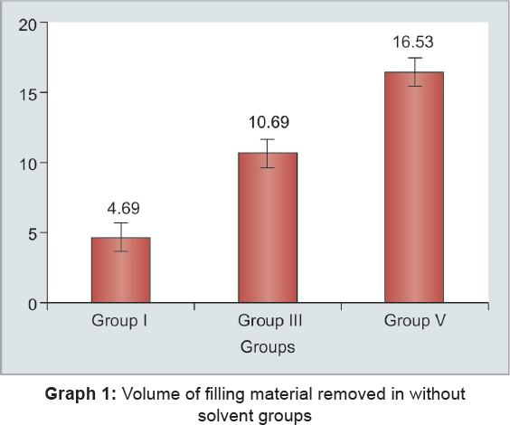
Table 1 presents the mean volume of filling material (in percentage) removed during retreatment, and the result shows that maximum percentage of filling material was removed by Rendo and ProTaper retreatment file with solvent and minimum percentage of filling material was removed by hand retreatment file with solvent, which is consistent with the finding of Gergi and Sabbagh.11
| Descriptive Statistics | ||||||
|---|---|---|---|---|---|---|
| Groups | n | Mean | Std. Deviation | Std. Error | Minimum | Maximum |
| 1 | 10 | 4.69 | 1.93 | 0.61 | 2.90 | 8.00 |
| 2 | 10 | 7.15 | 2.46 | 0.77 | 4.27 | 12.90 |
| 3 | 10 | 10.69 | 1.80 | 0.56 | 8.33 | 13.60 |
| 4 | 10 | 13.03 | 6.78 | 2.14 | 3.40 | 24.90 |
| 5 | 10 | 16.53 | 7.29 | 2.30 | 9.89 | 30.30 |
| 6 | 10 | 19.50 | 5.35 | 1.69 | 11.90 | 29.40 |
| One-way analysis of variance | ||||||
| Source of variation | Sum of squares | df | Mean square | f-value | p-value | |
| Between groups | 1565.03 | 5 | 313.007 | 13.32 | 0.0001, S < p < 0.05 | |
| Within groups | 1268.83 | 54 | 23.49 | |||
| Total | 2833.87 | 59 | ||||
In the present study all groups II, IV, VI when combined with solvent Endosolve R resulted in removal of more GP from canal walls as compared with nonsolvent group (Graphs 1 and 2). The mechanism of solvent in dissolving GP can be explained by their chemical properties. Gutta-percha is one of the natural rubbers, composed of trans-1,4-polyisoprene. The chemical principle, “like dissolves like,” means that a solute will dissolve well in a solvent that has a polarity similar to its own. For dissolving GP, a nonpolar solvent or less polar solvent must be used.12

The use of hand files was statistically different from the use of rotary instruments to remove filling material. This probably occurred because of GP plasticization resulting from rotation of the instrument.13 Softened GP is less resistant and easier to be penetrated and removed.
Under the experimental conditions of the present study, all techniques proved helpful for removal of endodontic filling material but complete removal of GP was not seen in any group, and there were no significant differences between rotary groups with solvent. But there was statistical significant difference seen between hand and rotary file systems without solvent.
The best removal of GP filling from canals was seen with R-Endo retreatment file with solvent followed by ProTaper retreatment file with solvent. Further studies are necessary to evaluate the effectiveness of this system in curved canals.
Source of support:
Nil
Conflict of interest:
None.
REFERENCES
- Endodontic retreatment-case selection and technique. Part 2. Treatment planning for retreatment. J Endod. ;1988J Endod. ;14(12):607-614.
- [CrossRef] [PubMed] [Google Scholar]
- Treatment outcome in endodontics: the Toronto study—phases 3 and 4: orthograde retreatment. J Endod. 2008;34(2):131-137.
- [CrossRef] [PubMed] [Google Scholar]
- Levels of evidence for the outcome of endodontic retreatment. J Endod. 2004;30(11):745-750.
- [CrossRef] [PubMed] [Google Scholar]
- The outcome of endodontic treatment: a retrospective study of 2000 cases performed by a specialist. J Endod. 2007;33(11):1278-1282.
- [CrossRef] [PubMed] [Google Scholar]
- The outcome of endodontic retreatment: 2 year follow-up. J Endod. 2004;30(1):1-4.
- [CrossRef] [PubMed] [Google Scholar]
- Comparative analysis of efficacy and cleaning ability of hand and rotary devices for gutta-percha removal in root canal retreatment: an in vitro study. J Contemp Dent Pract. 2013;14(4):635-643.
- [CrossRef] [PubMed] [Google Scholar]
- Efficacy of ProTaper universal retreatment files in removing filling materials during root canal retreatment. J Endod. 2008;34(11):1381-1384.
- [CrossRef] [PubMed] [Google Scholar]
- Efficacy of R-endo® and Protaper® re-treatment systems in removal of Realseal™. Aust J Basic Appl Sci. 2011;5(3):108-113.
- [Google Scholar]
- A removal of guttapercha and root canal sealer: a literature review and an audit comparing current practice in dental schools. Dent Update. 2012;39(10):703-708.
- [CrossRef] [PubMed] [Google Scholar]
- Removal efficiency of calcium hydroxide intra canal medicament with two calcium chelators: volumetric analysis using spiral CT–an in vitro study. J Endod. 2006;32(11):1097-1100.
- [CrossRef] [PubMed] [Google Scholar]
- Effectiveness of two nickel-titanium rotary instruments and a hand file for removing gutta-percha in severely curved root canals during retreatment: an ex vivo study. Int Endod J. 2007;40(7):532-537.
- [CrossRef] [PubMed] [Google Scholar]
- Efficacy of grapefruit, tangerine, lime, and lemon oils as solvents for softening gutta-percha in root canal retreatment procedures. J Investig Clin Dent. 2013;4(1):60-63.
- [CrossRef] [PubMed] [Google Scholar]
- Efficacy, cleaning ability and safety of different rotary NiTi instruments in root canal retreatment. Int Endod J. 2004;37(7):468-476.
- [CrossRef] [PubMed] [Google Scholar]


