

Translate this page into:
Results of the Pediatric Shaft Femur Fracture Treated with Closed Rigid Lateral Trochanteric Entry Intramedullary Nail in North Indian Population


Corresponding author: Dr. Siddharth Kothari, Department of Orthopaedics, Government Medical College and Hospital, Chandigarh, India. siddharthkothari11@gmail.com
-
Received: ,
Accepted: ,
How to cite this article: Kansay R, Vashisht S, Kothari S, Soni A, Jindal R, Garg S. Results of the Pediatric Shaft Femur Fracture Treated with Closed Rigid Lateral Trochanteric Entry Intramedullary Nail in North Indian Population. Int J Recent Sur Med Sci. 2023;9:105-10. doi: 10.25259/IJRSMS_1_2023
Abstract
Objectives
The femoral shaft fractures constitute 1.6% of the pediatric fractures. Intramedullary nailing is a common mode of fixation in older children and adolescents. The rigid intramedullary locking nail is a preferred choice over flexible nails in overweight patients and children older than 11 years of age. The adolescent age group with skeletal immaturity deserves special attention from the surgeons because of grave complications like avascular necrosis of the femur head and angular deformities. The aim of this study is to report the outcome of an adolescent femur shaft fracture treated with a rigid intramedullary interlocking nail in North Indian patients.
Material and Methods
This is a retrospective analysis performed by retrieving records of 19 patients. Patients with open fractures, polytrauma and neurovascular injuries were excluded. The preoperative and post-operative radiographic orthogonal views were analyzed to assess parameters like alignment, union and avascular necrosis (AVN).
Results
The mean age was 12.3 years. The mean follow-up was 1.5 years. The average time for union was 8 weeks. All the fractures were united in an acceptable alignment and rotation. There was no avascular necrosis, angular deformity and infection. The mean femur length discrepancy was 7 mm.
Conclusion
The rigid intramedullary interlocking nail is a valuable option for the fixation of femoral shaft fractures in an adolescent age group with a good union rate and minimal complications.
Keywords
Femur fracture
Adolescent
Rigid
Intramedullary nail
Skeletally immature

INTRODUCTION
Femur shaft fractures constitute 1.6% of overall fractures in children.[1] High-energy road traffic accidents are the commonest mode of injury in older children and adolescents, followed by sports injuries and falls from height.[2–4] The management of shaft femur fractures is age dependent. There is controversy and ongoing research regarding the gold standard of treatment for adolescent patients.[5] The treatment modalities include plating, minimally invasive plate osteosynthesis (MIPO), external fixation, elastic or rigid intramedullary nails.[2,6–8] Open reduction and internal fixation with plating result in extensive dissection, and subsequent scars also bother the patient. Moreover, there is an increased duration of restricted mobilization due to non-weight bearing.[1,9–13] External fixation is a more biological procedure and reduces hospital stay, but there are problems related to pin tract infection, and in some cases, refracture occurs after fixator removal.[14–16] The flexible intramedullary nail looks like an attractive option for surgeons, but there are some limitations. These include fracture comminution, obese children, fractures near the proximal or distal third, and limited rotational stability in adolescents.[14,17] The rigid interlocking nails provide adequate stability, are biology-preserving, and have good union rates.[6,18] However, there are some concerns regarding their use in children due to the occurrence of osteonecrosis of the femoral head or angular deformities of the proximal femur.[19,20] To avoid the dreaded complication of avascular necrosis (AVN), recent studies emphasized upon the lateral trochanteric entry point.[1,14] We describe the results of a lateral trochanteric entry rigid intramedullary nail in the pediatric adolescent patients having a femoral shaft fracture.
MATERIAL AND METHODS
This is a retrospective study performed at the orthopedics department of a tertiary care hospital. Institutional approval was obtained for including the patient in this study. The skeletally immature patients aged between 10 and 16 years, who had closed shaft femur fractures and were operated on between January 2016 and February 2018, were considered for this study. The records of operated patients were retrieved from the trauma database of the orthopedics department. Out of the 24 operated patients, the records of 19 patients with a minimum follow-up of 1.5 years were available for retrospective analysis. Patients with open fractures, polytrauma and neurovascular injuries were excluded. The preoperative and post-operative radiographic orthogonal views were analyzed to assess parameters like alignment, union and AVN. There were two patients who had been operated on previously at other institutes. There was bending of the flexible nail in one of them [Figure 1] and the other one had a broken implant with failure of fixation [Figure 2]. In both cases, the implant was removed and rigid nailing was done.
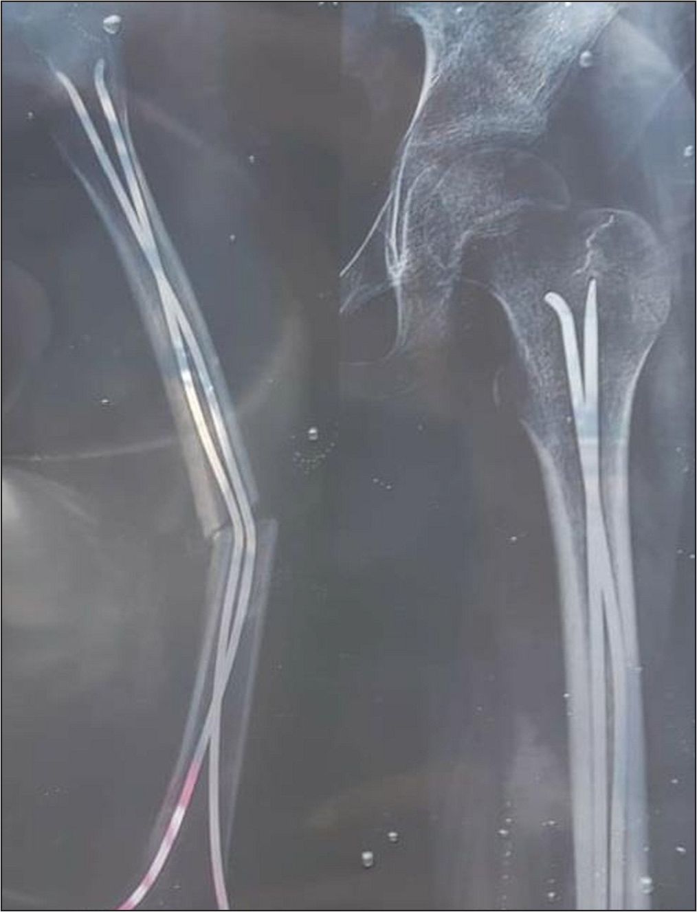
- Anteroposterior radiograph of a 12-year-old patient (50 kg) with bending of elastic nails.

- Anteroposterior radiograph showing failure of fixation with broken plate in an 11-year-old patient referred to our center.
TECHNIQUE
The patient was laid supine on the fracture table. After palpation of bony landmarks, a longitudinal skin incision (2 cm) was made over the tip of the greater trochanter (GT). Under C-arm guidance, the entry portal was made lateral to the tip of GT with the help of an awl. The guide wire was put inside, and an entry reamer was introduced. Closed reduction was achieved, and a guide wire was advanced into the distal fragment. Sequential reaming was done with a 1-mm increment in reamer size up to 9.5 mm. After confirmation of the nail length, a rigid intramedullary interlocking nail (Trigen adolescent nail [TAN] {Smith and Nephew, Memphis, TN, USA}) was put inside. After checking alignment and rotation, proximal and distal locking were done.
NAIL DESIGN
The intramedullary interlocking nail has a 12° lateral bend, which allows an entry point lateral to the tip of GT. The nail is made of titanium alloy and is side-specific. The nail diameter is 8.5 mm, and length varies from 26 to 40 mm in 2-mm increments. There is a single proximal lock and two distal locks. The distal-most lock provides a dynamic locking option.
Patients were gradually allowed progressive weight bearing from the second postoperative day with the help of a walker or crutches. In comminuted fractures, patients were allowed toe touch weight bearing.
RESULTS
A total of 19 patients were recruited [Table 1]. The mean age was 12.36 years. There were 12 male and seven female patients. 11 patients have right-sided injuries while eight patients have left-sided injuries. 89% of patients had road traffic accidents, and 11% of patients fell from heights. The mean follow-up was 1.5 years. The average time to union was 8 weeks (range: 6–13 weeks). The criteria for acceptable alignment include deviations up to 5° in the coronal and sagittal planes, and 10° in rotation. The neck shaft angle was measured and it was comparable to the normal side with no significant difference. All the fractures united in an acceptable alignment, and there was no case of nonunion or malunion [Figures 3–6]. The mean femur length discrepancy was 7 mm (range 5–11 mm). There was no infection. In one patient, the distal locking bolt was broken [Figure 7]. However, there was no need for reoperation for primary intervention. The fracture united and the broken bolt was removed. At the last follow-up (1.5 years), all the patients had normal hip and knee range of motion and normal gait. There was no pain at the fracture site or at the entry point. There was no clinical or radiological evidence of femoral head osteonecrosis in any case.
| Total patients (n) | 19 |
| Male: Female | 12:7 |
| Right: Left | 11:8 |
| Mean age (years) | 12.36 |
| RTA: Fall from height | 17:2 |
| Non union | 0 |
| Malunion | 0 |
| AVN | 0 |
| Deformity (varus/valgus) | 0 |
| Mean LLD (mm) | 7 |
RTA, road traffic accident: AVN, avascular necrosis; LLD, limb length discrepancy

- Preoperative radiograph of a patient with comminuted fracture of proximal third shaft femur.

- Postoperative radiograph of a patient with comminuted fracture of proximal third shaft femur showing union at the fracture site.

- Preoperative radiograph of a patient with long oblique fracture of shaft femur extending up to subtrochanteric region.

- Postoperative radiograph of a patient with long oblique fracture of shaft femur extending up to subtrochanteric region showing union at the fracture site.

- Postoperative radiograph showing complication in one case where distal locking bolt was broken. However, there is union at fracture site.
DISCUSSION
The management of femoral shaft fractures in older children and adolescents requires special attention for orthopedic surgeons. The two prime factors for deciding the optimal mode of fixation are canal diameter at the level of the isthmus and open physes.[5] The commonly performed procedures include intramedullary nailing, external fixation and plating. External fixators are preferred in open fractures, polytrauma and vascular injuries. The plating, especially MIPO, is preferred in cases where the canal diameter is very narrow. The intramedullary fixation methods have been minimally invasive and have provided good results in terms of union rates and complications.[21] Flexible nailing through the retrograde approach is preferred in the age group of 5–11 years. However, flexible nails are not suitable for long spiral and comminuted fractures, and these tend to bend in overweight children.[22–24] Other reported complications are irritation of the skin due to protruded nail ends, malunion and limb length discrepancy (LLD).[25] Ulici et al.[22] reported three prognostic factors that have influenced the outcome of the flexible nailing technique. These include age greater than 11 years, weight greater than 50 kg and the mode of injury. The last one was the most important and independent factor in the prediction of delayed union or deformity.
The rigid nails are preferred in patients with an age greater than 11 years, obese patients with a body weight greater than 49 kg, and unstable and comminuted fractures.
The femoral head derives its primary blood supply from the medial circumflex femoral artery. The branches of this vessel pierce the articular capsule at the level of the superior gemellus muscle. These small vessels finally ascend around the neck to supply the head region.[21,26] The proximity of this vasculature around the piriform fossa makes it vulnerable to disruption during entry point formation.[20,27,28] The entry point for antegrade nailing had been a matter of debate previously. The piriformis entry point had been disregarded in view of the reporting of a serious complication in the form of AVN. MacNeil et al.[29] performed a systematic review to find out the ideal entry point for skeletally immature patients. The three entry points – piriformis fossa, tip of GT and lateral aspect of the GT were compared. The lateral aspect of GT was found to be the safest entry point with the least risk to the vasculature around the hip. However, the coxa valga deformity had been reported in some studies where the nail passed either through the tip of the GT or some portion of the trochanteric physis.[30,31] The proximal femur develops from a single epiphysis in the early part of gestation. It then divides into two epiphyses (the capital femoral epiphysis and the GT apophysis) separated by an osseous portion. There is a remnant of epiphysis around the medial aspect of the GT and the lateral-most part of the neck. Gordon et al.[27] attributed injury to this specific portion, resulting in valgus deformity and narrowing of the neck. Therefore, the entry point at the lateral aspect of GT not only avoids the AVN but also prevents this valgus deformity. In our study, we used the lateral aspect of GT as an entry point, and none of the patients had AVN or the valgus deformity.
The comparison of studies reporting results of rigid intramedullary nails in the adolescent population is shown in Table 2.[32–34]
| Author | Journal | Year | Type of study | Total patients | Mean age (years) | Time to Union (weeks) | Union rate (%) | Entry site | Avascular necrosis | Infection | Mean Follow-up (years) |
|---|---|---|---|---|---|---|---|---|---|---|---|
| Townsend[32] | CORR | 2000 | Retrospective | 34 | 12.5 | NA | 100 | GT tip | 0 | 0 | 2 |
| Gordon[27] | JBJS | 2003 | Retrospective | 22 | 10.5 | NA | NA | Lateral GT | 0 | NA | 2 |
| Gordon[14] | JOT | 2004 | Retrospective | 15 | 12 | 7 | 100 | Lateral GT | 0 | 0 | 1.6 |
| Kanellopoulos[17] | JTIIC | 2006 | NA | 20 | 14.4 | 9 | 100 | GT tip | 0 | 0 | 2.4 |
| Mehlman[33] | JTIIC | 2007 | Retrospective | 6 | 12.5 | NA | 100 | GT | 0 | 0 | 14 |
| Keeler[1] | JPO | 2009 | Retrospective | 78 | 12.9 | 7 | 100 | Lateral GT | 0 | 2 | 1.1 |
| Elgohary[5] | EJOST | 2014 | Retrospective | 23 | 12.6 | 9 | 100 | GT tip | 0 | 0 | 2.5 |
| Chen[34] | JOT | 2018 | Retrospective | 112 | 10.3 | NA | 100 | GT | NA | 0 | 0.7 |
| This study | 2020 | Retrospective | 19 | 12.3 | 8 | 100 | Lateral GT | 0 | 0 | 1.5 |
CORR: Clinical Orthopedics and Related Research; JBJS: Journal of Bone and Joint Surgery; JTIIC: Journal of Trauma Injury, Infection, and Critical Care; JPO: Journal of Pediatric Orthopedics; EJOST: European Journal of Surgery and Trauma; JOT:Journal of Trauma; GT, Greater Trochanter; NA, not available.
All the previous studies had a retrospective design. The patient sample size ranged between 6 and 112 (mean 34.4). The mean age ranged from 10.3 to 14.4 years (average 12.2). The time to radiological union ranged between 7 and 9 weeks (average 8 weeks). There was complete union among all the patients. The GT was chosen as the entry point in all the cases. None of the cases had AVN of the femoral head. The mean follow-up ranged between 1.1 and 2.4 (average 1.6 years).
The main limitation of our study is its retrospective design. Moreover, there is no control group, and the sample size is small. High-quality randomized studies with comparison groups are required in the future.
CONCLUSION
The rigid intramedullary interlocking nail is a valuable option for the fixation of femoral shaft fractures in the adolescent age group with a good union rate and minimal complications.
Ethical Approval
The research/study complied with the Helsinki Declaration of 1964.
Declaration of patients consent
Patient's consent not required as patients identity is not disclosed or compromised.
Financial support and sponsorship
Nil.
Conflicts of interest
There are no conflicts of interest.
Use of artificial intelligence (AI)-assisted technology for manuscript preparation
The authors confirm that there was no use of artificial intelligence (AI)-assisted technology for assisting in the writing or editing of the manuscript and no images were manipulated using AI.
REFERENCES
- Antegrade intramedullary nailing of pediatric femoral fractures using an interlocking pediatric femoral nail and a lateral trochanteric entry point. J Pediatr Orthop. 2009;29:345-51.
- [CrossRef] [PubMed] [Google Scholar]
- Rigid intramedullary nail fixation of femoral fractures in adolescents: what evidence is available? J Orthop Traumatol. 2014;15:147-53.
- [CrossRef] [PubMed] [PubMed Central] [Google Scholar]
- Fractures of the femoral shaft in children. Incidence, mechanisms, and sociodemographic risk factors. J Bone Joint Surg Am. 1999;81:500-9.
- [CrossRef] [PubMed] [Google Scholar]
- Antegrade rigid nailing through the tip of the greater trochanter for pediatric femoral shaft fractures. Eur J Orthop Surg Traumatol. 2014;24:1229-35.
- [CrossRef] [PubMed] [Google Scholar]
- American Academy of Orthopaedic Surgeons clinical practice guideline on treatment of pediatric diaphyseal femur fracture. J Bone Joint Surg Am. 2010;92:1790-2.
- [CrossRef] [PubMed] [Google Scholar]
- Stabilization of pediatric diaphyseal femur fractures with flexible intramedullary nails (a technique paper) J Orthop Trauma. 1992;6:452-9.
- [CrossRef] [PubMed] [Google Scholar]
- Pediatric femoral fractures: a systematic review of 2422 cases. J Orthop Trauma. 2006;20:648-54.
- [CrossRef] [PubMed] [Google Scholar]
- Compression plating of pediatric femoral shaft fractures. J Pediatr Orthop. 2003;23:448-52.
- [PubMed] [Google Scholar]
- Compression plating for child and adolescent femur fractures. J Pediatr Orthop. 1992;12:626-32.
- [PubMed] [Google Scholar]
- Vascular injuries and amputation following limb fractures. Thorac Cardiovasc Surg. 1990;38:48-50.
- [CrossRef] [PubMed] [Google Scholar]
- Pediatric femur fractures: an overview of treatment. Orthopedics. 1993;16:183-90.
- [CrossRef] [PubMed] [Google Scholar]
- Management of pediatric femoral shaft fractures. J Am Acad Orthop Surg. 2004;12:347-59.
- [CrossRef] [PubMed] [Google Scholar]
- Intramedullary nailing of femoral fractures in children through the lateral aspect of the greater trochanter using a modified rigid humeral intramedullary nail: preliminary results of a new technique in 15 children. J Orthop Trauma. 2004;18:416-22; discussion 423–4.
- [CrossRef] [PubMed] [Google Scholar]
- Skeletal traction versus external fixation for pediatric femoral shaft fractures: a comparison of hospital costs and charges. J Orthop Trauma. 1998;12:563-8.
- [CrossRef] [PubMed] [Google Scholar]
- External fixation of femur fractures in children. J Pediatr Orthop. 1992;12:157-63.
- [CrossRef] [PubMed] [Google Scholar]
- Closed, locked intramedullary nailing of pediatric femoral shaft fractures through the tip of the greater trochanter. J Trauma. 2006;60:217-22; discussion 222–3.
- [CrossRef] [PubMed] [Google Scholar]
- Intramedullary nailing of pediatric femoral fractures. J Pediatr Orthop. 1994;14:184-9.
- [CrossRef] [PubMed] [Google Scholar]
- Intramedullary nailing of femoral fractures in adolescents. Clin Orthop Relat Res 1998:85-9.
- [PubMed] [Google Scholar]
- Avascular necrosis of the femoral head in an adolescent following intramedullary nailing of the femur. A case report. J Bone Joint Surg Am. 1994;76:1706-8.
- [CrossRef] [PubMed] [Google Scholar]
- Intramedullary nailing of pediatric femoral shaft fracture. J Am Acad Orthop Surg. 2011;19:472-81.
- [CrossRef] [PubMed] [Google Scholar]
- Poor prognostic factors of femoral shaft fractures in children treated by elastic intramedullary nailing. SICOT J. 2020;6:34.
- [CrossRef] [PubMed] [PubMed Central] [Google Scholar]
- Malunion following flexible intramedullary nails for tibial and femoral fractures in adolescents. J Child Orthop. 2010;4:571-7.
- [CrossRef] [PubMed] [Google Scholar]
- Biomechanical analysis of titanium elastic nail fixation in a pediatric femur fracture model. J Pediatr Orthop. 2008;28:874-8.
- [CrossRef] [PubMed] [Google Scholar]
- Complications of titanium elastic nails for pediatric femoral shaft fractures. J Pediatr Orthop. 2003;23:443-7.
- [PubMed] [Google Scholar]
- Trochanteric Entry for Femoral Lengthening Nails in Children: Is It Safe? J Pediatr Orthop. 2017;37:258-64.
- [CrossRef] [PubMed] [Google Scholar]
- Proximal femoral radiographic changes after lateral transtrochanteric intramedullary nail placement in children. J Bone Joint Surg Am. 2003;85:1295-301.
- [CrossRef] [PubMed] [Google Scholar]
- Femoral head avascular necrosis associated with intramedullary nailing in an adolescent. J Pediatr Orthop. 1995;15:21-3.
- [CrossRef] [PubMed] [Google Scholar]
- A systematic review of rigid, locked, intramedullary nail insertion sites and avascular necrosis of the femoral head in the skeletally immature. J Pediatr Orthop. 2011;31:377-80.
- [CrossRef] [PubMed] [Google Scholar]
- Intramedullary nailing of the femur in children. Effects on its proximal end. J Bone Joint Surg Br. 1995;77:262-6.
- [PubMed] [Google Scholar]
- Premature greater trochanteric epiphysiodesis secondary to intramedullary femoral rodding. J Pediatr Orthop. 1993;13:516-20.
- [CrossRef] [PubMed] [Google Scholar]
- Intramedullary nailing of femoral shaft fractures in children via the trochanter tip. Clin Orthop Relat Res 2000:113-8.
- [CrossRef] [PubMed] [Google Scholar]
- Tibial nails for femoral shaft fractures in large adolescents with open femoral physes. J Trauma. 2007;63:424-8.
- [CrossRef] [PubMed] [Google Scholar]
- Evaluation of Intramedullary Fixation for Pediatric Femoral Shaft Fractures in Developing Countries. J Orthop Trauma. 2018;32:e210-4.
- [CrossRef] [PubMed] [Google Scholar]


